
A wonderful review...even if you're not a student!
Written by Paul A. R. Meyer, a very impressive review of the slit-lamp was recently published in Nature: Eye. Whether your a seasoned clinician or not, this is a fantastic article celebrating the instrument we use everyday.
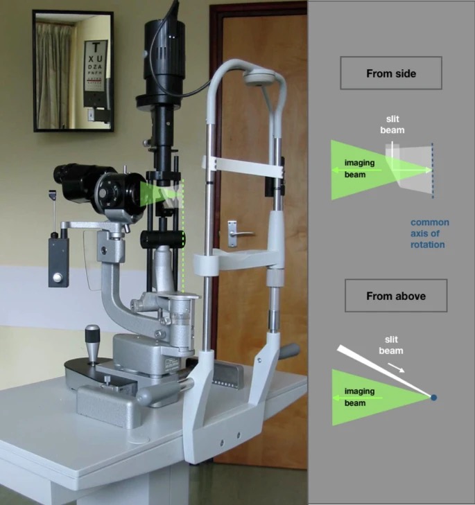
The slit-lamp biomicroscope was revolutionary by enabling detailed examination of ocular tissues "in-vivo". Combining a high-intensity light source that produces a thin, adjustable beam with a binocular microscope, it allows us to visualize eye structures with remarkable clarity.
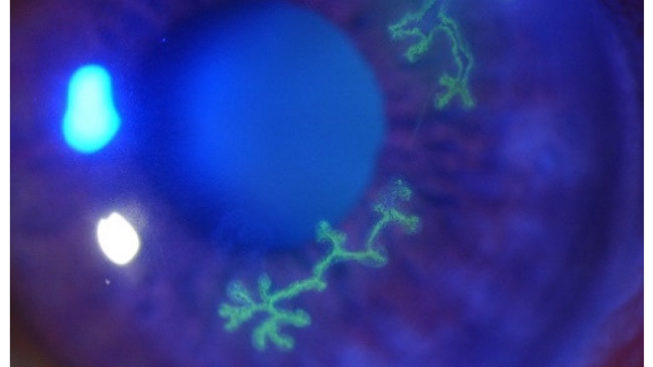
Key techniques include optical sectioning, which creates a cross-sectional view of transparent tissues, and retro-illumination, which utilizes reflected light to highlight subtle abnormalities. These methods permit detailed investigations of the cornea, anterior chamber, lens, and even portions of the vitreous and retina. By manipulating the angle, width, and intensity of the light beam, we can detect and characterize a wide range of ocular pathologies, from corneal ulcers to cataracts.
The slit-lamp's ability to provide "living histopathology" transformed ophthalmic diagnosis and management, keeping it an indispensable tool in modern eye care.
-JRM
Read the article in Nature: Eye...
Labs & Imaging For Primary Eye Care Course

- Downloadable Book & Companion Course
- Innovative "Go At Your Own Pace" Format
- 2 CE's & Instant Certificate
- Med-Eye Minute QAM Newsletter
- Powerful Learning Platform & VCA Community
HAVE A COURSE OR IDEA FOR A COURSE?
JOIN THE VIRTUAL COURSE ACADEMY!
READ THE BOOK SERIES ON AMAZON

Responses